
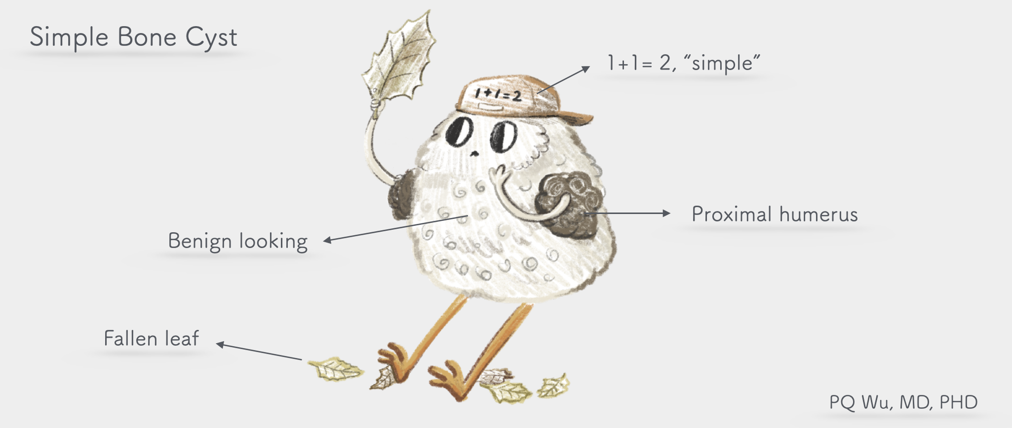
Simple bone cyst, a relatively common tumor, is mostly in adolescents younger than 20 years old. The main problem with the tumors is the cavity generated in the normal bones because of the regional growth issue.
Simple bone cysts are mostly undiscovered or have caused pathological fractures when they are found. Whether surgery is required depends on the tumor size, location, and patient symptoms. Overall, most of the cases require only tracking, not surgery.
Chinese: 單純性骨囊腫、孤立性骨囊腫
English: Unicameral Bone Cyst
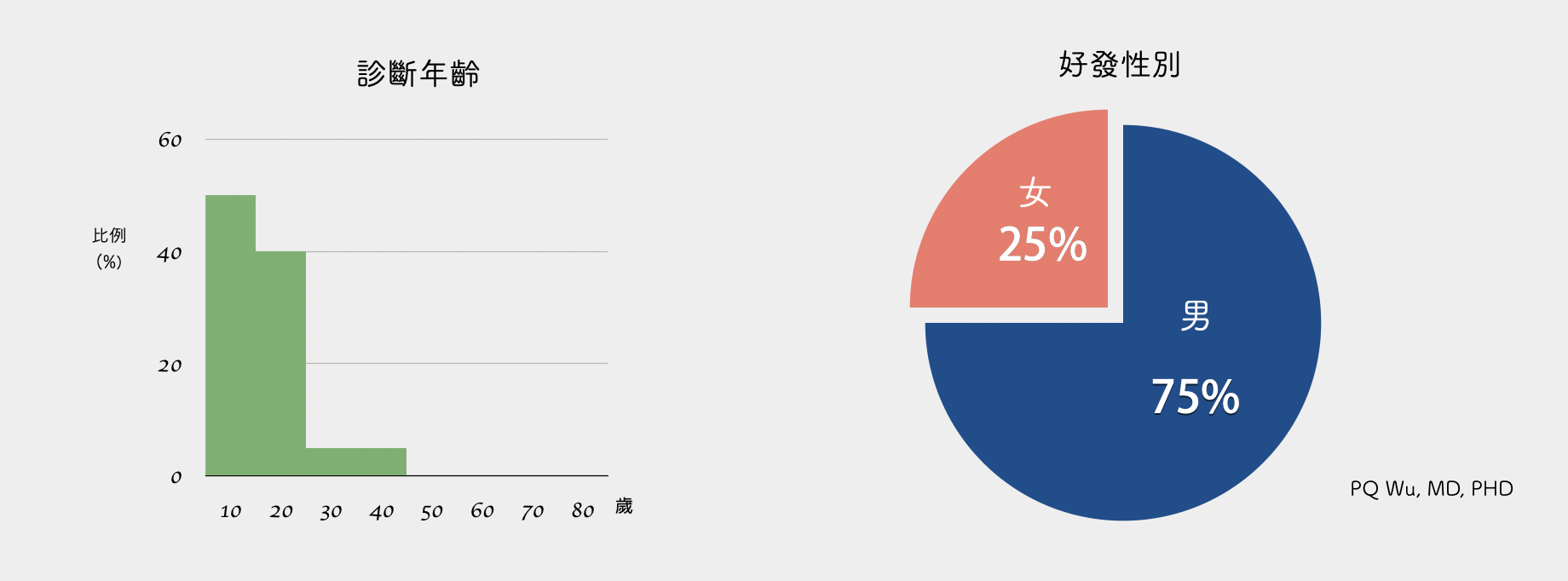
More than 80% of simple bone cysts occur in adolescents under the age of 20. If the patient is young, boys have a higher development chance than women, with a ratio of about 3:1. However, if it occurs at an older age (older than 20 years), the ratio of men to women is about 1:1.
Most simple bone cysts have no symptoms or are not even detected!
A small number of simple bone cysts are found after fractures
by PQ Wu
Most simple bone cysts are asymptomatic, and patients of this age (under 20 years of age) wouldn't conduct X-ray examination for no reason. Hence, most tumors are not detected. Occasional symptoms occur since the tumor is larger, and the patient comes to see the doctor for a sudden protruded piece of the appearance (especially at the proximal tibia and shoulder). However, most simple bone cysts are 'symptomatically' diagnosed by the pathological fractures that the patients suffer. Since boys of this age like to exercise and are very active, the common clinical situation is that the patient suffered the fracture in physical education class when he used his hand to touch the ground for support or fall and then was sent to the emergency room.
Simple bone cysts are common benign bone tumors. Since most cases are asymptomatic and stay steady with patients, medical diagnoses are usually not sought, and the correct incidence cannot be estimated. Our outpatient clinic would discover approximately 5 to 10 new patients in about one month at the Therapeutical and Research Center of Musculoskeletal Tumor in Taipei Veterans General Hospital.
Simple bone cysts would occur most commonly at the proximal humerus and then at the proximal femur. It is unlikely to see it in other locations. Since these two locations are common active and weight-bearing places, patients could suffer pathological fractures often. In addition, if the patient is older (older than 20 years old), a simple bone cyst would commonly occur at other rare locations such as calcaneus, iliac bone, and so on. If it occurs at the calcaneus, it often brings pain and pathological fractures to the patient because the location is the weight-bearing place for walking and running
Simple bone cyst mostly originated from the metaphysis, and it is almost unlikely to invade epiphysis and joint surface. Medically, we divide the bones into epiphysis, metaphysis, and diaphysis. Most bone tumors occur at the epiphysis

Simple bone cysts have a rather special feature. The tumor that grows at the metaphysis will grow 'higher', pushed by the newly developed bone from the metaphysis to the diaphysis as the patient ages.
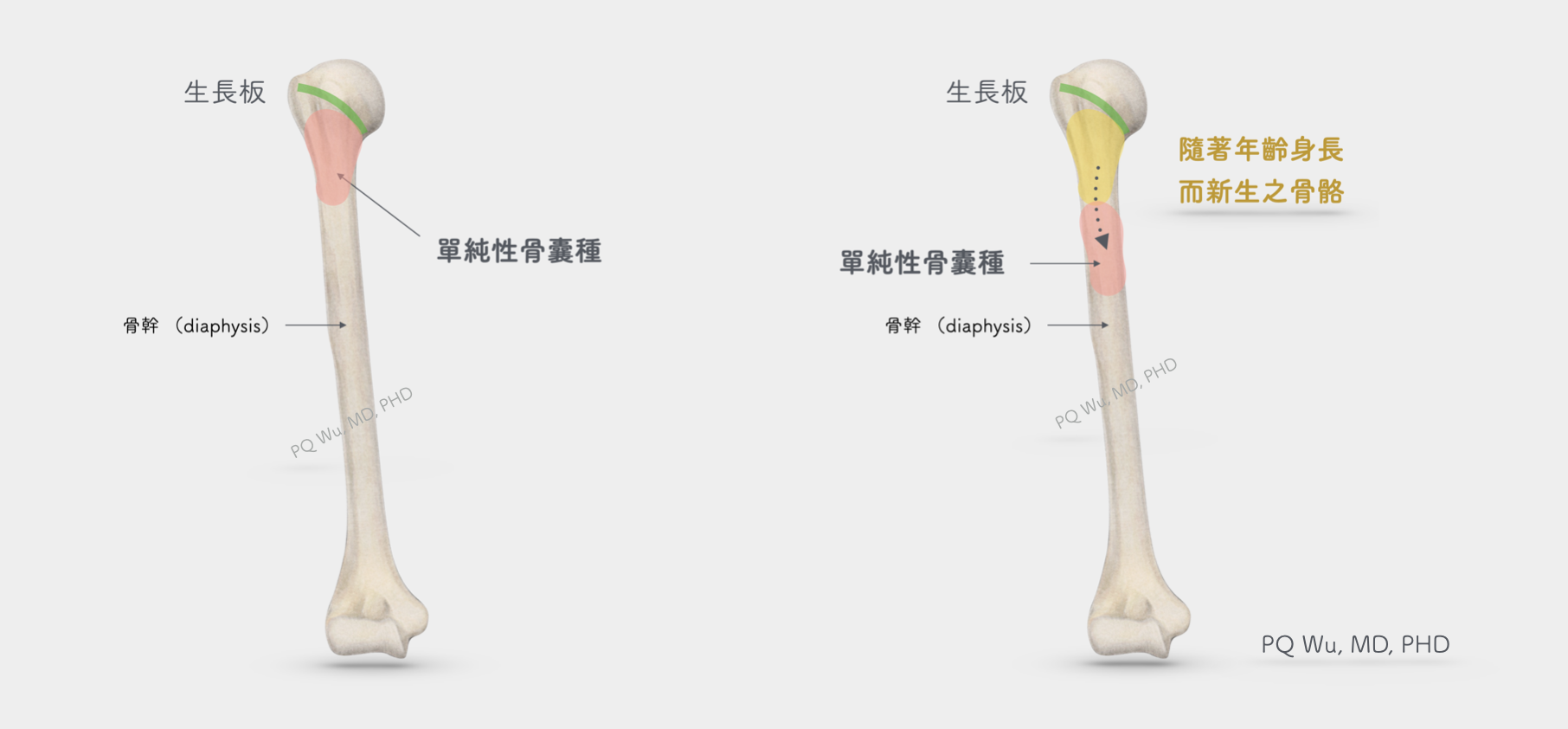
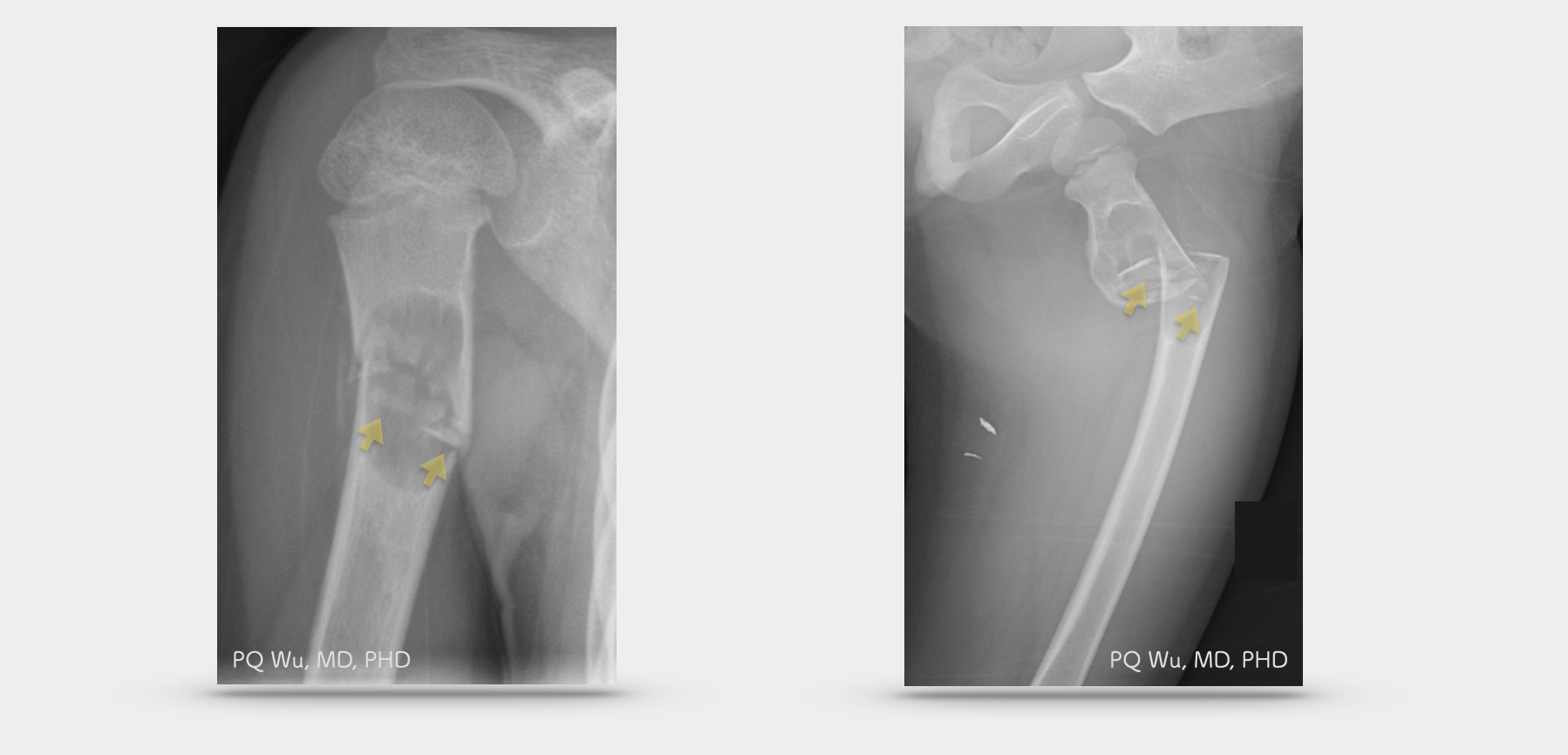
When the patient is young, the simple bone cyst grew at the metaphysis (upper left). As the bone lengthens, the simple bone cyst was pushed to the diaphysis (upper right)
Under X-ray images, a simple bone cyst grows more in the central location of the bone, generating mild expansion of the bone. Unless there is a pathological fracture, the periosteal reaction will not occur. It also won't break out of the bones and invade the surrounding soft tissue. In MRI images, homogeneous intermediate signal intensity in the T1 signal exists at the lesion, and homogeneous high signal intensity in the T2 signal is discovered. These are typical discoveries of simple bone cysts.
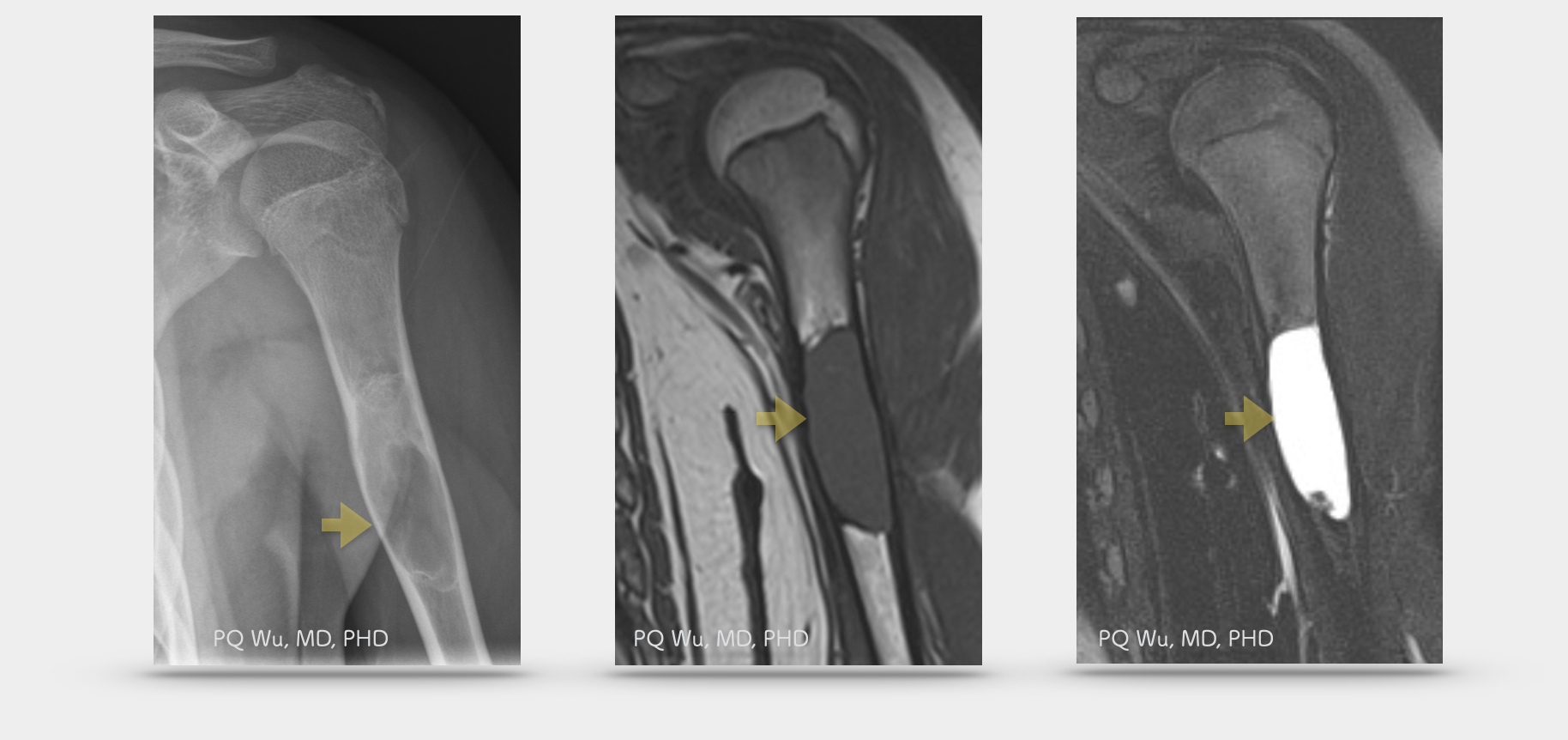
X-ray image of the expanded bone resulted from a simple bone cyst (upper left). MRI image (T1) with a homogeneous intermediate signal intensity (upper middle). MRI image (T2) with a homogeneous high signal intensity (upper right).
A ten-year-old girl with a simple bone cyst combining with pathological fractures (upper left). A four-year-old boy with a simple bone cyst combining with pathological fracture (upper right).
The cause of simple bone cysts is still unknown. Most studies suggest that simple bone cysts are not tumors but merely dysplastic or reactive lesions than true tumors. The causes are thought to be:
1. Calcification problem of the cartilage in the growth plates
2. In cancellous bone, venous circulation problems that lead to local pressure rise, triggering inflammatory reactions and bone absorption. (*,**)
* Tumeur osseuses bénignes. Paris: Elsevier; 2005. p. 176–87.
**KaelinA.Kystesessentielsdesos.In:DuparcJ,editor.Cahiersd’enseignementsde la Sofcot. Paris: L’Expansion scientifique franc ̧ aise; 1995. p. 167–79
Pathological diagnosis of simple bone cysts must rely on the 'exclusion method.' In appearance, when we surgically enter a simple bone cyst, we see clear water with a little yellow serum-like fluid. In the process of scraping the tumor, a few fibrocystic-like structures will reveal. If we see this phenomenon in surgery, we can directly determine it as a benign simple bone cyst. But in some cases, because simple bone cysts will combine pathological fractures, the cyst structure will bleed and become fibrosis, and the liquid outflow will appear bright red with blood. The surgeon will be quite troubled at this time! Such a phenomenon will make it difficult to distinguish it from benign aneurysmal bone cysts, giant cell tumors, and even malignant telangiectatic osteosarcoma. So in surgery, if we find something unusual like this, i.e., findings during surgery are different from the preoperative images, experienced physicians will decide to minimize the wound and scope to avoid contamination. Followed by waiting for a few days for the formal pathology report to come out, they proceed to the next step. Otherwise, bad things will happen
Simple bone cysts are single and will not grow everywhere!
by PQ WuSimple bone cysts basically won't be multiple. Sporadic cases of multiple simple bone cysts appear with the chance of less than 1%.
Simple bone cysts basically won't turn malignant!
by PQ Wu
Simple bone cysts won't turn malignant; however, as mentioned earlier, since it's not easy to diagnose, the location difference of tumor specimen sampling would result in a misdiagnosis that other malignant tumors be diagnosed as simple bone cysts.
Most simple bone cysts do not require surgical treatment.
However, if the tumor locates close to the proximal tibia that is near the shoulder or to the femur (near the hip), the tumor range is relatively large, and the bone structure becomes more porous that causing pathological fractures easily, we will generally recommend surgical treatment to avoid subsequent complications.
The procedure includes two steps: tumor removal and bone transplantation in the lesions. I'll detailly describe these two steps in the following articles. You can access it through the links below.
In addition, because simple bone cysts are likely to heal automatically, we can also make a hole in the bone that generates a smaller wound so that the fluid inside the tumor can flow out to achieve the purpose of decompression. While this method is simpler, the likelihood of recurrence is higher, and it takes longer for the bone to heal since bone transplantation is not introduced. Therefore, we use this method less often in our treatment protocol at the Therapeutical and Research Center of Musculoskeletal Tumor in Taipei Veterans General Hospital.
Regular post-surgical outpatient tracking is required. The doctor will determine how much force you can step on and when you can be free from crutches based on how well your bones recover. After full recovery, there's a chance of recurrence of fibrous dysplasia, and regular X-ray checks will still be needed so as to detect and treat problems early.