
Chondroblastoma occurs mostly in younger patients. Because where it occurs is on the joint surface, it often causes more serious effects on limb function. Overall, chondroblastomas are quite similar to giant cell tumors in adults. Although they are essentially benign bone tumors, they have large local damages to bones, are prone to recurrence, and sometimes even incur lung metastasis. These characteristics are a headache for orthopedic surgeons, but now because of the advance in diagnostic imaging and surgery, these problems can be found and treated early!
Chinese: None
English: Codman tumor
Most patients experience pain. If the symptom persists for a period of time, muscle atrophy and limited joint movement can result from the fear of taking force at the lesion.
Chondroblastoma is a relatively rare benign bone tumor, taking about 9% of all benign bone tumors and 1% of all bone tumors. An outpatient clinic would discover a new patient in about three months at our Therapeutical and Research Center of Musculoskeletal Tumor in Taipei Veterans General Hospital. It is relatively uncommon in all benign bone tumors.
Chondroblastomas are about twice as likely to occur in men as in women. Most tumors occur in children and adolescents between the ages of 5 and 20, and generally, they rarely occur in adults more than 18 years old whose growth plates are closed. This feature is the opposite of giant cell tumors.
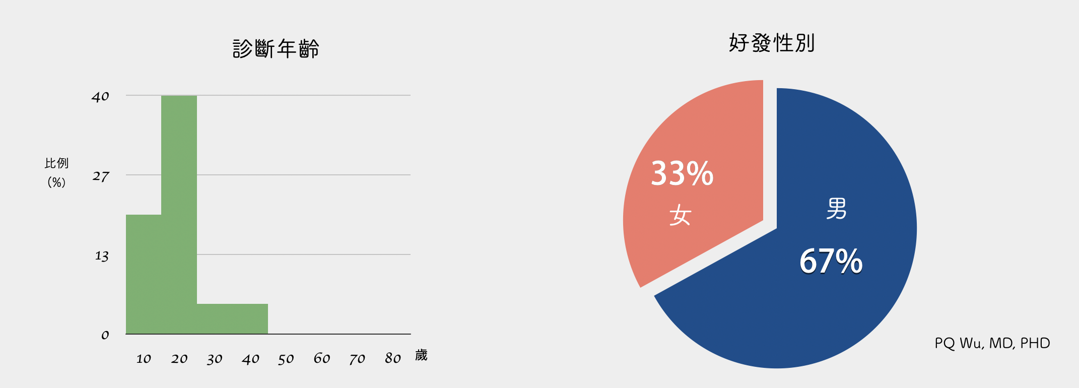
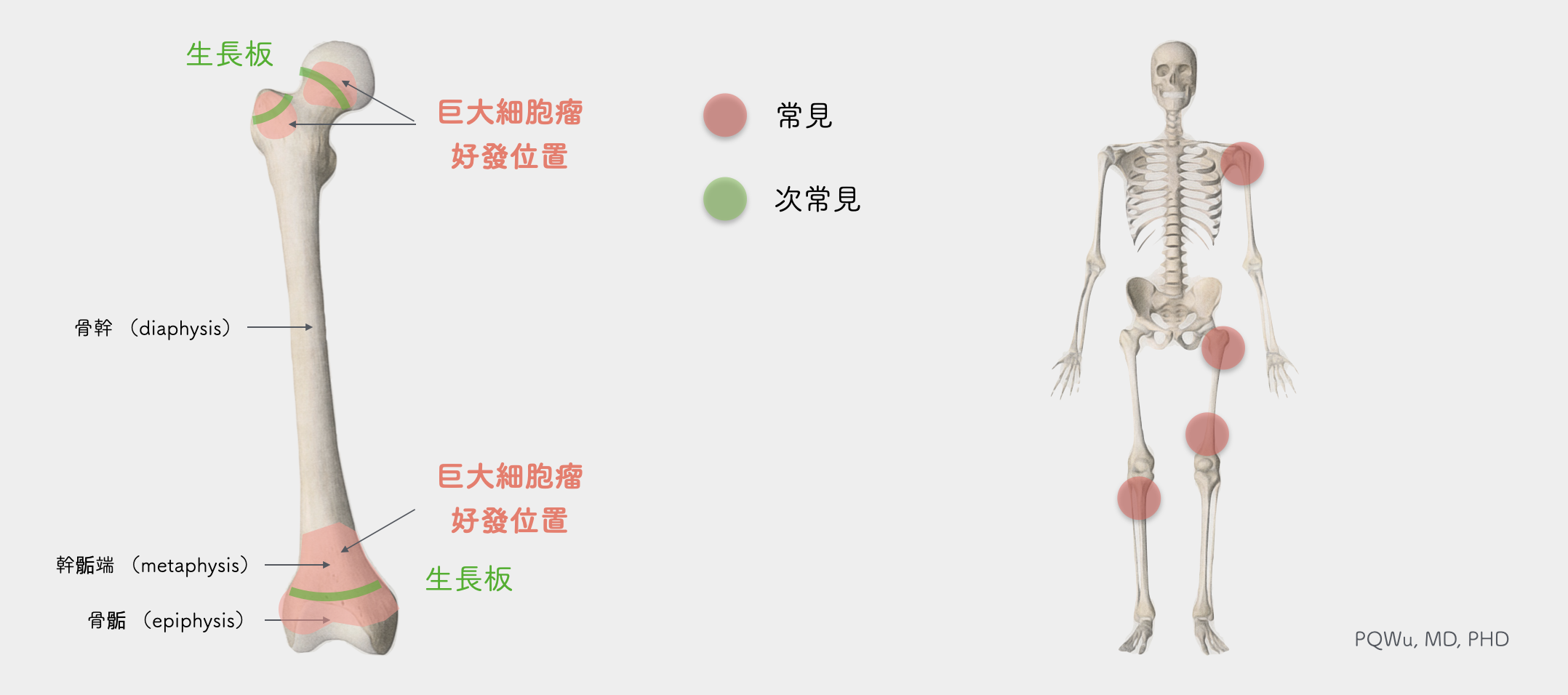
Chondroblastomas mostly occur in the distal femur, proximal tibia (above and below the knee), and proximal humerus.
Most bone tumors occur mostly at the metaphysis, while chondroblastoma and giant cell tumor are two exceptions. Chondroblastomas occur most likely at the epiphysis near the joint. Tumors that grow at the epiphysis are less easy to have surgical treatments, and chondroblastomas tend to bring great distress to physicians since they occur in patients whose growth plates have not yet been closed. Damage to the growth plates resulting in uneven leg length would be the concern if we want to remove the tumor thoroughly. However, to reduce the damage of the growth plate, the tumor often can not be excised thoroughly and will likely to recur in the future. Also, since the tumor is closer to the joint surface, the impact on patients' limb activity will be greater if post-surgical rehabilitation is not enforced. (Medically, we divide the bones into epiphysis, metaphysis, and diaphysis.)
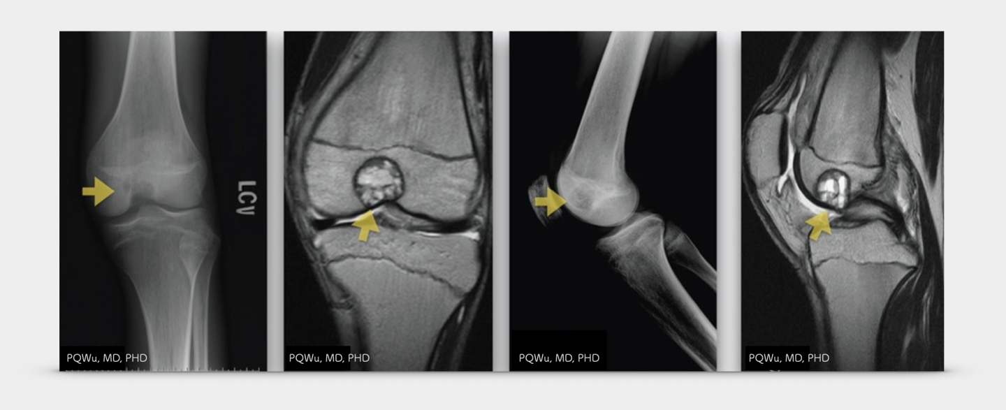
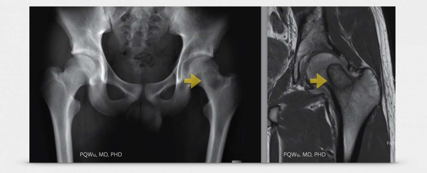
Chondroblastomas in X-ray image exhibit osteolytic lesions at the bone of epiphysis and metaphysis near the joint. The lesions are usually more towards one side and have thin sclerotic borders. The overall appearances reveal geographic patterns, and most of them are round or egg round, which is quite special. About half of chondroblastomas will have calcification. In the MRI image, nearby soft tissues and bone marrow edema are obvious. Hence, it is very easy to be mistakenly recognized as a malignant tumor. Let's take a look at the image examinations of several patients:
Currently, we have a limited medical understanding of the cause of chondroblastoma! In 1998, the formation of chondroblastoma was found associated with the gene translocation of the fifth and eighth pairs of chromosomes at the University of Nebraska Medical Center*. Unfortunately, there are still no further new discoveries.
*Significance of abnormalities of chromosomes 5 and 8 in chondroblastoma.Clin Orthop Relat Res. 1998 Apr;(349):189-93.
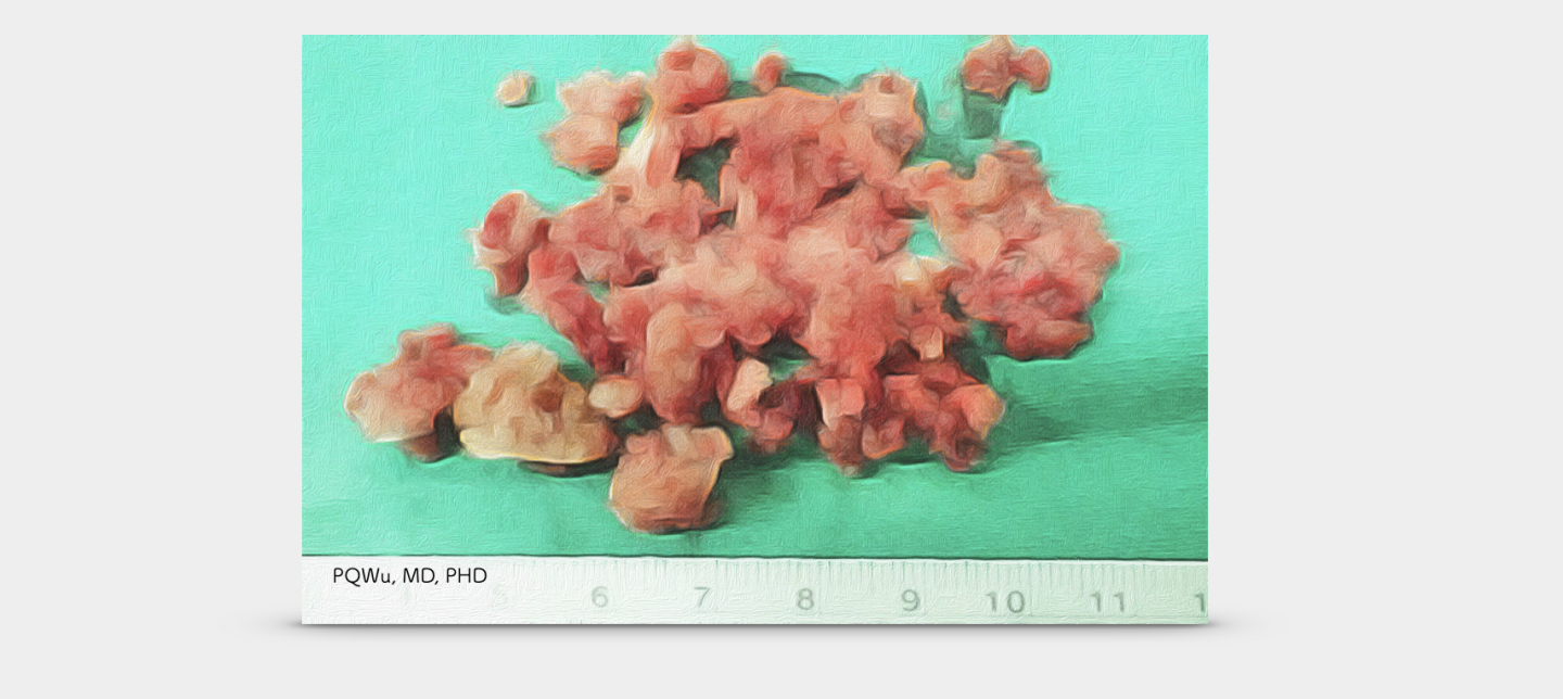
Chondroblastoma has a soft pattern when we touch, and its overall color is white, combining bleeding and festering.
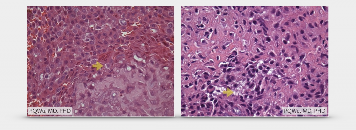
Under the microscope, the cell pattern of chondroblastomas is unique. Long circles like pebbles are medically referred to as "cobblestones." In addition, there are many scattered giant cells, or osteoclast-like Giant cells, in tumors. Giant cells are bone-breaking cells that may be attracted by tumor matrix cells. There are about 20 or more nuclei in the cell. The mitotic phenomenon is often seen, but there is no atypical mitosis, which is the leading cause of bone damage via chondroblastoma. In some cases, we can see calcification between cells. Medically, we'd call it as "chicken-wire calcification", or "Lattice-like-matrix calcification" since it looks like the chicken coop with hexagonal mesh wire.
Although chondroblastoma is benign, 1% of lung metastasis occurs and needs to be tracked regularly!
Although chondroblastoma is a benign tumor, in the general literature, chondroblastoma can be found to have a 1% chance of lung metastasis (same as it for giant cell tumors). Regular lung examinations are also necessary
* Chondroblastoma with pulmonary metastasis in a patient presenting with spontaneous bilateral pneumothorax: Report of a case.Surg Today. 2011 Oct;41(10):1439-41
**Benign chondroblastoma. Case report with pulmonary metastasis.J Bone Joint Surg Am. 1975 Apr;57(3):418-20
***A case of spine origin chondroblastoma metastasis to lung.Cancer Res Treat. 2009 Dec;41(4):241-4. doi: 10.4143/crt.2009.41.4.241. Epub 2009 Dec 31.
One percent of chondroblastoma, although benign, will turn into malignant. It'd need attention, albeit the chance is very low!
by PQ WuOnly two cases that the chondroblastoma turned into malignant were reported in medical journals(*,**), and both of them had fairly low chance. However, what annoyed physicians are that some chondroblastomas performed more aggressively than tumor cells, and it often causes serious local bone damage, multiple relapses, and a quite poor limb recovery! It even turns into lung metastasis!
Another rare case of chondroblastoma was an osteoblastoma-like OGS, which performs as same as normal chondroblastoma in the initial phase and grows rapidly after surgery or tracking for a period of time, generating serious problems!
* Malignant transformation of chondroblastoma.Histopathology. 1996 Nov;29(5):477-80.
**Benign chondroblastoma of bone. Report of a case of malignant transformation. J Bone Joint Surg Br 1970;52:741–745.
Surgery for chondroblastoma is absolutely necessary! Surgeons will need surgery to remove the tumor and then transplant the bones to fill in the bone defects. However, chondroblastoma is fairly easy to recur. If we only remove the tumor via scraping, the chance of recurrence after surgery as high as 32.5%. So, the adjuvant therapy during surgery is very important.
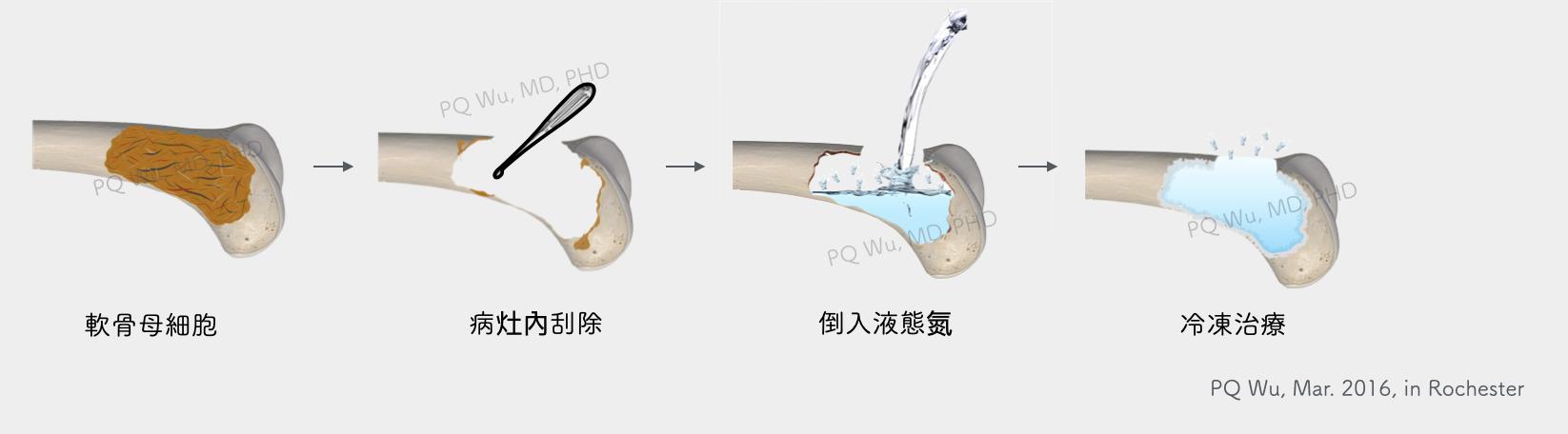 Therefore, three surgical steps for the chondroblastoma removal, including tumor removal in the lesions, adjuvant therapy after surgery, and bone transplantation, are detailed in the following articles below: