
When we highly suspect that a patient has osteosarcoma, several imaging examinations are required. These include X-ray scan of the affected region, MRI scan of the affected region, chest CT scan, whole body bone scan, and whole abdominal sonogram. Although PET is quite accurate, it's not widespread since currently it's not reimbursed by the National Health Insurance and it's relatively costly. In terms of blood tests, unfortunately there is no specific tumor marker to monitor osteosarcoma. Therefore, we can only use indirect serum ALK-P and LDH indices to understand the severity of the disease.
X-ray scanning are the most convenient and the most important test to diagnose osteosarcoma.
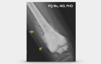
An experienced osteosarcoma specialist can tell almost 80 percent of osteosarcoma with a typical osteosarcoma X-ray image. Osteosarcoma has several (but not absolute) characteristics under X-rays, including:
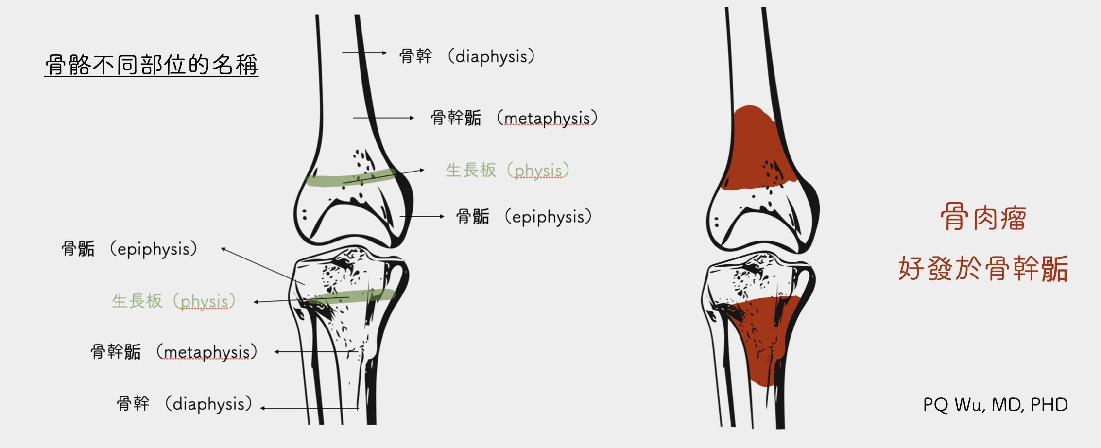
1. Commonly occurring at the metaphysis, occasionally invading the physis and entering the epiphysis.
2. Osteoblastic, osteolytic, or mixed lesions appear on the bones.
3. Codman triangle or sunburst periosteal reaction appear on the periosteum.
Descriptions for each characteristics on X-ray are as follows:
I. Commonly occurring at the metaphysis, occasionally invading the physis and entering the epiphysis.
Medically, we devided the bone, from the joint end to the far end, into epiphysis, metaphysis, and diaphysis. Most of osteosarcomas grow at metaphysis because of the distribution of blood vessels. Osteosarcoma, along with most of other bone tumors, occurs at the metaphysis. Of course, if the tumor is discovered later, or if the tumor itself grows faster, the chance of osteosarcoma invasion to metaphysis and diaphysis is still quite high.
2. Osteoblastic, osteolytic, or mixed lesions appear on the bones.
These osteosarcomas differs considerably under X-rays. Since tumor cells in the bones will induce many biohormones, promoting the growth of osteoblast and osteoclast in nearby regions. Under normal physiological conditions, osteoblasts promote bone regrowth, and osteoclasts destroy old bones. With the cooperation between the two cells, a normal bone metabolism is achieved. Under osteosarcoma conditions, however, the balance is disrupted. Hence, in some cases, osteoblasts continue to abnormally promote bone growth, resulting in so-called Osteoblastic Osteosarcoma. Conversely, in some cases, osteoclasts are constantly and abnormally to break down bones, generating so-called Osteolytic Osteosarcoma. In addition, some osteosarcomas have osteoblastic and osteoclastic elements, which we call Hybrid Osteosarcoma. The readers can click on the link below for detailed descriptions on mechanism of bone destruction by tumor cells.
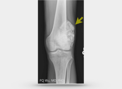
Osteoblastic osteosarcoma
In the X-ray image, many abnormal bone structures in tumor can be clearly observed. The bone structure in osteoblastic tumor is stronger and less likely to incur pathological fractures.
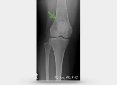
Osteolytic osteosarcoma
In the X-ray image, obvious cavities on bone at the tumor site can be clear viewed. Therefore, the bone structure of osteoclastic osteosarcoma is very fragile and prone for pathological fractures.
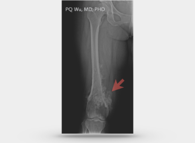
Hybrid Osteosarcoma
In the X-ray image, both osteoblastic and osteoblastic elements in the tumor can be clearly telled.
3. Codman triangle or sunburst periosteal reaction appear on the periosteum
As mentioned above, the abnormal bone regrowth and destruction can also occur on the periosteum, resulting in different shapes of periosteal reaction. In general, as long as we see these such as triangular, reticular, or sunburst periosteal reactions, we can determine that the tumor grows rapidly, and the chance of being malignant is quite high.
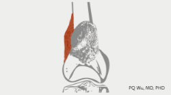
Codman triangle periosteal reaction
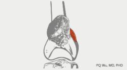
Reticulum periosteal reaction
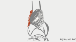
Sunburst periosteal reaction
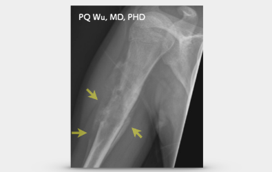
Codman triangle periosteal reaction
Sunburst periosteal reaction
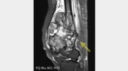
MRI can precisely locate the range of osteosarcoma invasion so that it gives orthopedists a clear idea of how long a bone needs to be removed. In addition, it can also give us a clear understanding of the relationship between osteosarcoma and those important normal tissues or organs nearby, such as nerves, blood vessels, ligaments, tendons, skin, and abdominal organs. If these important tissues or organs are found to be invaded or severely contaminated by tumors, orthopedists will need to collaborate with other surgeons such as colorectal surgery, urology surgery, cardiovascular surgery and plastic surgery specialists. In addition, in recent years, the radiation department team (Dr. HT Wu, Dr. CL Wu) in our Therapeutical and Research Center of Musculoskeletal Tumor has been able to use imaging information (DWI, Diffusion Weight Image) to determine the tumor necrosis rate after chemotherapy. This not only provides considerable helps to our surgeons in surgery but also provides an absolute reference for oncologists to choose chemotherapy drugs.
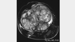
MRI can accurately find the relationships among tumor, nerves, and blood vessels
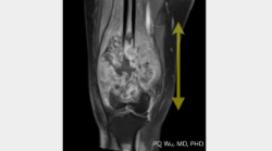
MRI can accurately measure the length of the tumor
MRI can accurately find the relationship among tumor, nerves, and blood vessels
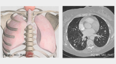
Lung is the location where metastasis is most likely to occur, and once metastasis occurs, it has a considerable impact on the overall prognosis. Generally, smaller lung lesions are unlikely found under X-ray; therefore, it's necessary to use chest CT for lung tracking.
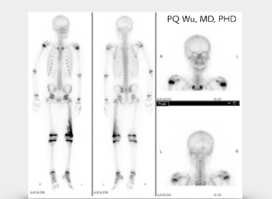
The chances of bone metastasis are not high in the early stages of osteosarcoma. However, at initial diagnosis and follow-up stage, whole body bone scans are still required to identify the occurrence of bone metastasis.
Whole body bone scan can be used to precisely determine whether lung metastasis occurs
For other medical centers, lesion sonograms are mostly used only as initial screening and examination of tumors. However, it's not just that in Therapeutical and Research Center of Musculoskeletal Tumor at Taipei Veterans General Hospital. Dr. HR Chiou, the director and the "National Treasure" in our center, can also use ultrasound to detect the tumor blood flow so that he can identify the rate of tumor malignancy and necrosis. The accuracy of Director Chiou's judgment before surgeries often makes us orthopeadists astonished. He deserves the name "National Treasure"!
The chances of metastasis to abdominal organs, such as liver, are low in osteosarcoma. However, at initial diagnosis, whole abdominal sonogram is still required to identify the occurrence of abdominal organ metastasis.
Serum ALK-P stores in human bones. When bones are destroyed and regenerated (e.g. in bone cancer, fractures, etc.), the blood concentration of serum ALK-P increases significantly. In our studies of some other osteocarcinomas, we found that if the patient's serum ALK-P value is higher, a poor prognosis is implied. However
LDH is an enzyme for glucose metabolism. When cells in the body are destroyed, the amount of the LDH in blood increases. In our studies of some other osteosarcomas, we found that if patients had higher LDH values, higher chances of developing lung metastasis are implied. However, some studies found no correlation between the two.
切片檢查相當重要,是診斷骨肉瘤最重要的項目。我會在下一篇文章詳細說明。
The examinations mentioned above, including imaging and blood drawing, are important examinations before or after osteosarcoma diagnosis. The results of these tests often influence the course of treatment because they have a considerable relationship with the prognosis of the disease. Hence, it is important to complete these tests completely and track them regularly!